Cornea
- Home
- Cornea
Contact lenses are thin, clear plastic disks you wear in your eye to improve your vision. Contacts float on the tear film that covers your cornea.


Cornea Overview
Cornea is the clear outer layer of the eye. A number of conditions affect the cornea that leads to “Corneal disease”. The cornea can often repair itself after injury or disease, but more serious conditions — infections, degenerative diseases, deterioration — need treatment.
Individuals with corneal blindness are usually of a younger age group compared with those suffering from cataract. Hence, in terms of total blind years, the impact of corneal blindness is greater.
In India, ocular trauma, infectious keratitis, corneal ulceration and post-infectious keratitis corneo-iridic scars contribute significantly to ocular morbidity in children.
In adults, the major causes of corneal blindness include bacterial, fungal or viral keratitis, hereditary corneal dystrophy and eye injuries. The major causes of corneal morbidity in the elderly include trachomatous keratopathy, corneal degenerations and trauma-induced infectious keratitis.
Common Causes of Corneal Disease

Infection
Bacterial, fungal and viral infections are common causes of corneal damage

Corneal Ulcers
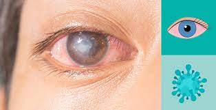
A corneal ulcer is a small sore that grows on your cornea. The cornea is the clear tissue that covers the front part of your eye. In most cases, an infection, injury or wearing contact lenses (contact ulcers) causes a corneal ulcer.
Other Risk Factors Causing Corneal Ulcers
Chronically dry eyes, weakened immune systems, and seasonal or chronic allergies, you are also more prone to developing corneal ulcers.
Aging processes can affect the clarity and health of the cornea.
Bullous keratopathy occurs in a very small percentage of patients following these procedures.
- Heredity
- Eye trauma
- Systemic diseases, such as Leber’s congenital amaurosis, Ehlers-Danlos syndrome
- Down syndrome and osteogenesis imperfecta.
symptoms of corneal diseases are:
- Pain
- Blurred vision
- Tearing
- Redness
- Extreme sensitivity to light
- Corneal scarring.
our services
What to Expect at Sankar Foundation
At Sankar Foundation, we are equipped to treat all types of corneal diseases:
- Congential and hereditary corneal disorders
- Corneal and scleral infections
- Ectatic corneal disorders – Keratoconus, Terriens marginal degeneration, Pellucid Marginal degeneration
- Immunologic disorders of cornea
- Peripheral ulcerative keratitis
- Ocular surface disorders including severe dry eye, blepharitis, ocular allergies, chemical injuries, Stevens Johnson syndrome and ocular surface tumors.
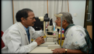
We have some of the most advanced diagnostic equipment for treating corneal conditions.
Evaluates shape and power of the corneal surface and is able to obtain the accurate measurement of elevations, curvature, power and thickness for the whole cornea.
Live microscopic imaging of corneal disorders
Measurement of corneal thickness using ultrasound.
( I-Trace Aberrometer, Allegro Analyser) Measures the optical aberrations of the eye.
Invaluable tool in dry eye workup , meibography.
Measures tear osmolarity helping detect dry eye.
Photographic documentation of various corneal conditions for baseline documentation, and monitoring effects of therapy as well as to monitor progression of the disease in any ectatic disorder.
Corneal Transplant
A cornea transplant is performed to improve the function of the cornea and improve vision. An eye donated is used to replace the diseased cornea.
Sometimes through injury, infection, or some inherited condition (such as keratoconus)
cornea may lose its natural transparency or normal shape, leading to blurred vision.
In many cases glasses or contact lenses may help you to see more clearly, but there are times when
these do not work and the cornea needs to be replaced (graft/transplant).
Cornea transplant is a procedure that replaces your cornea, the clear front layer of your eye. During this procedure, your surgeon removes damaged or diseased corneal tissue. Healthy corneal tissue from the eye of a deceased human donor replaces the damaged cornea. For many people, cornea transplant surgery restores clear vision and improves their quality of life.
Congenital corneal disorders are also an important cause of childhood blindness that usually result from hereditary dystrophies, congenital glaucoma, Peter’s anomaly and other mesenchymal dysgenesis, birth trauma and metabolic disorders.
A Rare Surgery by Dr Nasrin
At the age of 13, Maka Ganga Nageswara Rao suffered an eye injury. While eating sugarcane, particle of the sugarcane struck his right eye, turning red and painful developing into a corneal ulcer. He went to several primary and secondary care government hospitals for treatment but did not meet with success and he decided to live with the problem.
After 17 long years, in 2010, he attended a camp by Sankar Foundation. After a thorough evaluation by Ophthalmologists at Sankar Foundation, he was diagnosed as having Adherent Leukoma with Cataract in Right Eye. A Penetrating Keratoplasty along with iOL implantation surgery was planned. After thorough counselling, Dr. Nasrin successfully performed this difficult and rare surgery on the 5th of May, 2010.
After the surgery, Maka was a ‘visibly’ happy man as there was a perceptible improvement in his vision. It was a long delayed but ‘ sweet recovery for him. When he was explained by the Counsellors of the Foundation that the surgery was possible because somebody has donated his eyes, Maka felt very touched. He promised himself and to Sankar Foundation that he will actively propagate the idea of Eye Donation’ so that more people like him who are suffering from avoidable corneal blindness can benefit and lead a happy life.


 Smt. K. Mani Mala is a postgraduate holding an MBA. She started her career as a founder faculty of one of the first management colleges in Visakhapatnam, viz. Indian Institute of Advanced Management. She worked with East India Petroleum Limited as its first employee and as Executive Assistant to the Managing Director. She was also associated in establishing the Joint Venture with M/s SHV Energy of Netherlands as General Manager (MIS). During her tenure she was also associated with other group companies such as 16 MW & 450 MW gas-based power projects in AP and 16 MW & 100 MW hydel power plants in Himachal Pradesh.
 ( Late) Dr. Kumar studied Law and Economics at the Andhra University, Visakhapatnam and Chennai. He was a Chartered Accountant of repute with offices in Visakhapatnam, Hyderabad and Chennai.